Are you suffering from blurred vision? Or are you seeing sudden flashes of light that appear when you look on the sides and floaters inside the eyes? These are all the symptoms of retinal detachment. To understand retinal detachment in detail, one needs to learn what is retina. The retina is a layer at the back of the eyeball and is located near the optic nerve. Retina receives light from the lens, converts it into neural signals and sends these to the brain for visual recognition. Though there are numerous issues that occur in the retina, retinal detachment is one of the most talked-about eye disorders.
What is retinal detachment?
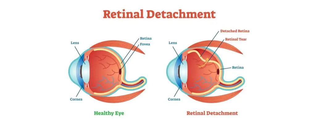
It is a detachment of a thin layer of retina from the layer of blood vessels that provides it with nutrients and oxygen. In this eye condition, a break in the retina allows the fluid in the eye to go behind the retina. Retinal detachment disorder leads to worsening of the outer part of the visual field. It is like a curtain over a part of the field of vision. If left untreated, a retinal detachment which is also known as detached retina can result in permanent vision loss.
Symptoms of retinal detachment
- Blurred vision
- If you notice sudden spots, floaters (black or grey specks that seem to float away when the person tries to look directly at them) and flashes of light, then these are the warning signs too
- Partial vision loss. This vision loss would feel like that a curtain has been pulled across the field of your vision. You may experience a shadow effect too
- The detached retina is not usually painful
There are mainly three types of retinal detachment. Let’s understand all of them:
- Rhegmatogenous: This is considered to be the most common type of retinal detachment. In this type, a tear occurs in the retina. The tear allows fluid from within the eye to get into the back of the retina. The area where retina detaches lose the obvious blood supply and discontinue to work, leading to loss of vision.
- Tractional: In this type of retinal detachment, scar tissue starts to grow on retina’s surface, eventually pulling retina from the back of the eye. Tractional retinal detachment is typically seen in people who have diabetes.
- Exudative: This kind of retinal detachment is different from Rhegmatogenous retinal detachment in a way that in this eye condition, fluid accumulates beneath the retina. There are no tears in the retina. Causes of this can be aging, injury to the eyes, inflammatory disorders, etc.
Treatment for Retinal Detachment
Treatment for this eye disorder includes laser eye surgery, freezing treatment and other types of surgery depending upon the intensity with which retinal detachment has occurred. Initially, the doctor will check the vision, physical appearance of the eye, eye pressure and how eyes are reacting seeing different colors. An eye ultrasound can also be done. Doctors may check the ability of the retina to send impulses to the brain. They may analyze the flow of blood to the retina. Some of the detached retina treatments include:
- Retinopexy: In this way of treatment, doctors put gas bubbles to support the retina move back to its place against the wall of your eye.
- Cryopexy: In this procedure, doctors apply freezing probe outside the eyes, the scarring takes place because it holds retina backs to its place.
- Vitrectomy: This option is used in patients with larger tears. Doctors may need small tools to remove scar tissue and vitreous from retina. Then they put the retina back to its place with gas bubble.
- Scleral Buckling: In this procedure, doctors put a band outside the eyes in order to push the wall of the eye into the retina.
If you have been looking for a trusted eye care partner who could help you in getting healthy eyes then get connected with Centre For Sight . CFS is a leading eye care provider in India which provides eye care solutions to various complex eye problems.


