Papilledema is a medical condition characterized by swelling of the optic disc, the area where the optic nerve connects to the eye. It is typically caused by increased pressure in the brain transmitted to optic nerves, which can be a result of various underlying conditions. This blog post will provide an overview of papilledema symptoms, causes, and treatment options.
This condition can affect individuals of all ages, although it is more commonly observed in adults. Prompt diagnosis and papilledema treatment are essential to prevent potential complications and preserve vision.
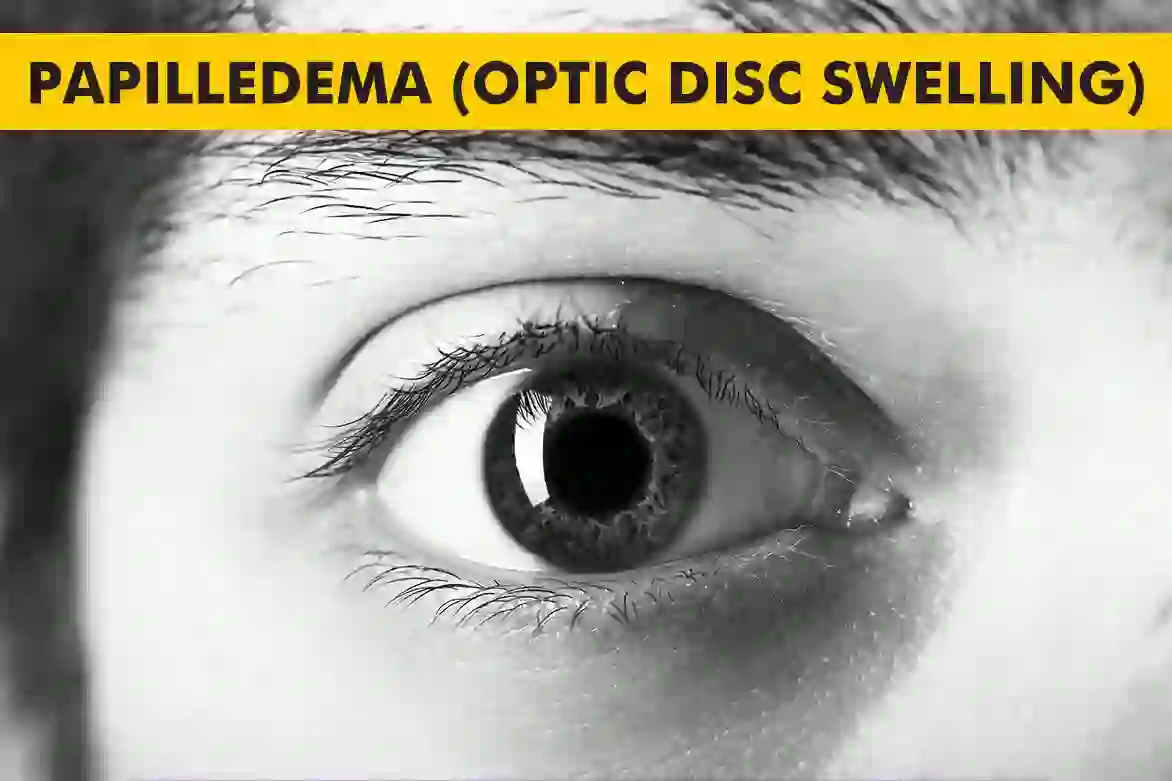
What Is Papilledema?
To understand papilledema definition, it is important to first have some knowledge of the optic disc. The optic disc is the spot on the retina where the optic nerve enters the eye. It contains blood vessels and nerve fibers that transmit visual information to the brain. In the presence of increased intracranial pressure, the optic disc can become swollen, resulting in papilledema.
Papilledema is different from optic disc edema, which refers to the swelling of the optic disc due to other causes such as inflammation or infection. Papilledema specifically indicates optic disc swelling due to increased pressure in the brain and usually affects both the optic nerves simultaneously.
The exact mechanisms behind papilledema are not fully understood. However, it is believed that the increased pressure in the brain leads to impaired drainage of cerebrospinal fluid, causing it to accumulate and exert pressure on the optic disc. This pressure then leads to the swelling and subsequent changes in vision.
Know more about the high eye pressure here.
Types of Papilledema
There are two main types of papilledema: bilateral and unilateral.
Bilateral papilledema refers to optic disc swelling in both eyes. This is the most common type and is often associated with conditions such as intracranial hypertension, brain tumors, or meningitis.
Unilateral papilledema, on the other hand, involves optic disc swelling in only one eye. This can be caused by conditions such as optic neuritis, anterior ischemic optic neuropathy, or local venous congestion.
In general, papilledema refers to bilateral involvement of the optic nerve.
The Different Papilledema Stages
Papilledema stages can be classified into different levels based on the severity of optic disc swelling and associated changes. These papilledema stages help in assessing the progression of the condition and determining appropriate papilledema treatment strategies. The stages of papilledema are as follows:
Stage 0: No visible swelling of the optic disc, but other signs of increased intracranial pressure may be present.
Stage 1: Mild swelling of the optic disc, with some elevation and blurring of its margins.
Stage 2: Moderate swelling of the optic disc, with more pronounced elevation and blurring of its margins.
Stage 3: Severe swelling of the optic disc, with marked elevation and loss of normal disc landmarks.
Stage 4: End-stage papilledema, characterized by optic disc swelling accompanied by hemorrhages and exudates.
It is important for healthcare professionals to accurately assess papilledema stage to determine the appropriate course of treatment.
Signs and Symptoms of Papilledema
Papilledema is often asymptomatic in its early stages, making it difficult to detect without a comprehensive eye examination. However, as the condition progresses, various papilledema symptoms may manifest, including:
- Blurred vision
- Vision loss
- Headaches
- Nausea and vomiting
- Seeing flashing lights or halos around lights
- Difficulty with peripheral vision
Here are the types of low vision you should know.
If any of these papilledema symptoms are experienced, it is important to seek medical attention promptly to prevent further damage to the optic nerve and potential vision loss.
Causes of Papilloedema
Papilledema is primarily caused by increased pressure within the skull, known as intracranial pressure. Several conditions and factors can contribute to this increased pressure, including:
- Brain tumors
- Meningitis
- Encephalitis
- Hydrocephalus
- Idiopathic intracranial hypertension
- Cerebral venous sinus thrombosis
- Traumatic brain injury
These conditions can lead to the accumulation of cerebrospinal fluid or the growth of abnormal tissues, resulting in increased pressure on the optic disc and subsequent papilledema.
Diagnosis and Evaluation
Diagnosing papilledema involves a comprehensive evaluation by an ophthalmologist or neurologist. The diagnostic process may include the following:
- Visual acuity test: Assessing the clarity of vision.
- Visual field test: Evaluating the extent of peripheral vision.
- Dilated eye examination: Examining the optic disc for signs of swelling.
- Optical coherence tomography (OCT): Producing cross-sectional images of the retina and optic nerve.
- Lumbar puncture (spinal tap): Measuring the pressure of cerebrospinal fluid.
These diagnostic tests help in confirming the presence of papilledema and determining its underlying cause. Additional imaging studies, such as magnetic resonance imaging (MRI) or computed tomography (CT) scans, may also be conducted to identify any structural abnormalities within the brain.
Treatment for Papilledema
The primary goal of papilledema treatment is to reduce intracranial pressure and prevent further damage to the optic nerve. The specific treatment approach will depend on the underlying cause and severity of the condition. Possible papilledema treatment options include:
Medications: Diuretics or other medications may be prescribed to reduce fluid buildup and lower intracranial pressure.
Surgery: In certain cases, surgical intervention may be necessary to alleviate the underlying cause of papilledema, such as removing a brain tumor or repairing an obstructed cerebral vein.
Shunting procedures: Some individuals with papilledema may require the placement of a shunt, a device that helps divert excess cerebrospinal fluid to another part of the body where it can be absorbed.
Vision therapy: In cases where vision loss has occurred, vision therapy may be recommended to help individuals adapt and make the most of their remaining vision.
It is important for individuals with papilledema to follow their healthcare provider’s recommended treatment plan and attend regular follow-up appointments to monitor their condition.
Complications and Prognosis of Papilledema
If left untreated, papilledema can lead to serious complications and permanent vision loss. The increased intracranial pressure can cause damage to the optic nerve, resulting in irreversible vision impairment.
However, with early diagnosis and appropriate treatment, the prognosis for papilledema can be favorable. By effectively managing the underlying cause and reducing intracranial pressure, the swelling of the optic disc can be alleviated, and vision loss can be prevented or minimized.
Regular monitoring and follow-up appointments with healthcare professionals are essential to ensure the effectiveness of the treatment and prevent potential complications.
Preventive Measures of Papilledema
While it may not always be possible to prevent papilledema, certain measures can help reduce the risk or severity of the condition. These preventive measures include:
Managing underlying conditions: Effectively managing conditions such as intracranial hypertension, brain tumors, or infections can help prevent the development of papilledema.
Regular eye examinations: Routine eye examinations can help detect any early signs of optic disc swelling or other eye conditions.
Healthy lifestyle choices: Maintaining a healthy lifestyle, including a balanced diet, regular exercise, and proper sleep, may help reduce the risk of developing conditions that can lead to papilledema.
It is important to consult with healthcare professionals for personalized advice and guidance on preventive measures based on individual risk factors and medical history.
Summary
Papilledema indicates a space occupying lesion in the skull or brain & hastily identifying the cause can prevent irreversible damages.
An exact determination will require various tests, alongside the contribution of eye and mind subject matter experts. These people can decide how best to deal with the condition.
FAQs
What is the main cause of papilledema?
Main cause of papilledema is increased intracranial pressure (ICP) that affects the optic nerve.
What is the treatment for papilledema?
Addressing the underlying cause, often involving medications to reduce intracranial pressure or surgery in some cases.
What are the 4 stages of papilledema?
Stages of papilledema: Mild, moderate, severe, and chronic (advanced).
Is papilledema a brain tumor?
Papilledema can be caused by brain tumors, but it is not exclusive to tumors; it can result from various conditions that raise intracranial pressure.
Can papilledema cure itself?
No, papilledema typically requires treatment to resolve. It can worsen if not managed.
Which vitamin deficiency causes papilledema?
Vitamin A deficiency can lead to papilledema.
Is papilloedema life threatening?
Papilledema itself is not life-threatening but can be vision threatening. It indicates a serious underlying condition (such as a brain tumor or severe intracranial pressure increase) that can be life-threatening if not treated promptly.