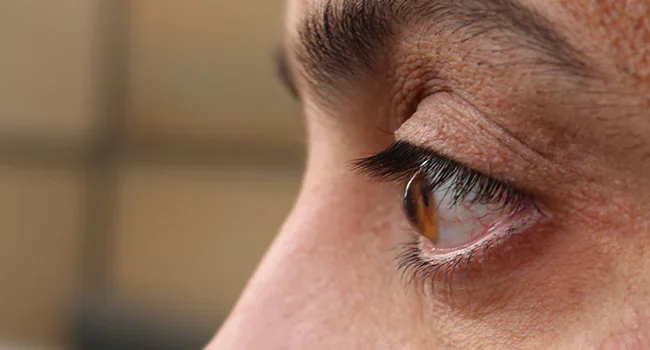
Keratoconus is a progressive disease that affects the patient’s cornea. The cornea, the top transparent layer of our eyes, gets thin and slopes down due to the progressive disease keratoconus (cone-like). As a result, the light rays entering the eye don’t focus on a single point, leading to hazy and distorted vision. Patients may notice shadows or duplication of pictures. Keratoconus can worsen and impair vision significantly if neglected. Usually, it affects both eyes differently, and the early signs are most frequently seen in the late teenage years.
Four types of Keratoconus
The types of Keratoconus are determined by the cornea’s shape and the area where the cornea has thinned. In addition to uncommon variations, the disease comes in four main types.
Nipple cone keratoconus or round keratoconus
Although just a tiny portion of the cornea is affected by round or nipple cone keratoconus, the slope of the afflicted area can be extremely steep. This kind of Keratoconus causes a noticeable decline in vision.
Sagging cone keratoconus or oval keratoconus:
A wider area of the cornea is impacted by oval or sagging cone keratoconus. The internal membranes of the cornea may tear (or hydrops), leading to scarring and making contact lens fitting more challenging.
Forme fruste keratoconus
The most popular form of Keratoconus is called fruste. In actuality, former fruste Keratoconus is typically symptomless and can only be identified through mapping the eye’s surface. Forme fruste keratoconus is a condition that first manifests but does not proceed for whatever reason. Treatment is not usually necessary for those who have formed fruste Keratoconus.
Keratoglobus keratoconus
A unique form of Keratoconus causes the entire cornea to thin and extend forward. The optimum vision correction typically includes using conjunctival lenses with a big diameter.
Signs and symptoms of keratoconus
Nearsightedness is a result of Keratoconus (myopia). This implies that you have problems perceiving distant objects. Astigmatism is also a result of it. This is an issue with the retina of your eye focusing on a picture. All of this results in vision blurs.
Typically, symptoms begin in teenagers and worsen until about the age of 40. If your eye doctor doesn’t do specific testing, you might not even be aware that you have this disorder. Your vision could become significantly worse later. If your vision deteriorates faster than predicted, your doctor might examine you for Keratoconus.
Different Keratoconus Stages
Here we discuss the stages of Keratoconus-
1. Initial Keratoconus:
Only very tiny corneal deformation, little to no impact on vision quality, and little to no progression are all characteristics of early-stage Keratoconus. Spectacles typically provide adequate eyesight while successfully treating myopia and astigmatism.
2. Moderate Keratoconus:
In this stage, it is possible to see corneal changes typical of Keratoconus and increased corneal distortion. Rigid gas permeable contact lenses are an alternative for enhanced vision as the quality of vision with glasses declines.
3. Advanced Keratoconus:
At this stage, there is significant corneal deformation, mild keratoconus alterations, and light to severe corneal scarring. The rigid gas permeable contact lens design may be subject to more modifications than for moderate Keratoconus, frequently utilizing much higher interior curvatures to maintain an effective fitting.
4. Severe Keratoconus:
Dramatic corneal deformation, significant corneal scarring, and thinning are all symptoms of severe Keratoconus. With stiff gas permeable contact lenses, eyesight is frequently poor, contact lens tolerance is significantly reduced, and it’s typically quite challenging to properly fit a rigid gas permeable contact lens. If we talk about severe keratoconus treatment, it is advised to contemplate a corneal transplant after being referred to a skilled corneal surgeon.
How to identify keratoconus?
Your eye doctor will examine your eyes and inquire about your medical background. They will assess your vision’s clarity. You might need to dilate your eyes for a portion of the exam. Your healthcare professional will prescribe eye drops to you to enlarge (dilate) the black area in the center of your eye. This enables your provider to get a clearer view of more of your eye. The shape of your cornea may also be measured by a tool by your healthcare professional. Your doctor might use a corneal topography imaging test to aid in the diagnosis. Changes in the cornea’s shape are visible with this examination.
Treatment for keratoconus
The course of your keratoconus treatment may change depending on how severe your Keratoconus is. Your specific kind of Keratoconus may also affect your keratoconus treatment options.
Early on, you might require glasses to correct the keratoconus-related vision impairment. If glasses don’t work, another alternative is to use specialized contact lenses. These connections are frequently gas-permeable. These need to be fitted carefully to your cornea.
Many patients won’t require any additional care. However, you could need a different treatment if your cornea becomes too damaged or unable to support a contact lens. A corneal transplant is the standard course of action in this case. Part or all of the native corneal thickness is removed with this procedure. It is changed with a deceased donor’s cornea (cadaver donor).
There are a variety of Keratoconus treatments, such as:
- Collagen Crosslinking in the Cornea (CXL)
- contact lens (Custom Soft, Gas Permeable, Rose K, Corneal, Scleral, and Mini-scleral)
- Implants by Intacs
- Implantation of Personalized Stromal Lenticules
- Photorefractive keratectomy (PRK) with topography guidance and CXL
- Deep anterior lamellar keratoplasty for corneal transplantation (DALK).
- Why Choose CFS?
As one of India’s most well-known eye hospitals, Centre for Sight, takes pleasure in providing the best Keratoconus treatment. We are dedicated to providing our patients with fresh perspectives and cost-effective solutions. C3R or CXL, also known as corneal collagen crosslinking with riboflavin, is a promising treatment for Keratoconus.
Centre for Sight is well-equipped with cutting-edge technology and ultra-modern technology for the early evaluation and treatment of Keratoconus.
Article: Keratoconus Eye Disorder: Types, Signs, & Treatments
Author: CFS Editorial Team | Sep 16 2022 | UPDATED 04:27 IST
*The views expressed here are solely those of the author in his private capacity and do not in any way represent the views of Centre for Sight.
