|
Key Takeaways:
|
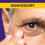
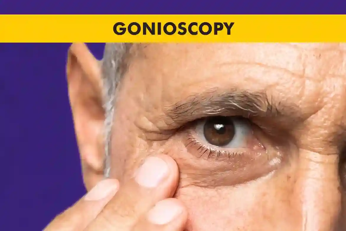
Gonioscopy is a simple eye test that lets your doctor see the drainage angle where the cornea and iris meet, an area that is not visible in a routine check-up. Many people hear the term and feel anxious because they don’t know the gonioscopy meaning, what the lens does, or whether the test will hurt.
In this article, you’ll learn what is gonioscopy, how the gonioscopy procedure is done step by step, what the doctor looks for in the angle, and how gonioscopy grading helps in diagnosing and managing glaucoma.
Gonioscopy Meaning
To understand the gonioscopy meaning, it helps to first know what doctors are looking at inside the eye. At the front of the eye, the clear window (cornea) meets the coloured part (iris) and forms a tiny angle, this is called the drainage angle.
Here, a clear fluid called aqueous humor leaves the eye through a natural filter and then drains into the bloodstream, helping to keep eye pressure in a healthy range.
In a healthy eye, this angle is open and the fluid flows out smoothly.
If the angle is narrow, blocked, or built abnormally, the fluid cannot leave easily, eye pressure may rise, and over time this can damage the optic nerve and cause glaucoma. Gonioscopy is a special microscope-based gonioscopy test that lets the eye doctor directly see this drainage angle, check its shape, and look for any scars, pigment, or abnormal vessels.
This information is essential for diagnosing and managing different types of glaucoma and for planning the right treatment.
Gonioscopy Structures
During a gonioscopy procedure, your ophthalmologist looks closely at the fine details of the drainage angle. By studying how clearly these structures are seen, and whether anything is blocking them, your doctor can judge how safely the fluid can leave the eye.
These gonioscopy structures include:
- Trabecular meshwork: A sieve-like tissue at the base of the cornea that acts as the main drain, allowing aqueous humor to filter out of the eye.
- Schlemm’s canal: A circular channel just behind the trabecular meshwork that collects this fluid and passes it into tiny veins.
- Iris root: The place where the iris joins the eye wall and helps decide how open or narrow the angle is.
Other landmarks like the Schwalbe’s line and ciliary body band, which help in gonioscopy grading of how “open” or “closed” the angle appears.
Why Is Gonioscopy Necessary?
Gonioscopy is a key test whenever your doctor is worried about glaucoma or angle problems. It helps them find out whether the drainage angle is open, narrow, or already closed, and whether there are any abnormal blood vessels, pigment, or scar tissue in this area. This is important because two eyes can have the same pressure but very different angle findings, which may need different treatments.
The gonioscopy test is especially useful for spotting eyes at risk of angle-closure glaucoma, where the angle can shut suddenly, causing a rapid rise in pressure, severe pain, and blurred vision. If the angle looks narrow or shows signs of previous closure, your doctor can advise preventive laser treatment before an attack happens.
Gonioscopy is also used in conditions like uveitis, trauma, or developmental problems of the front of the eye to see how the angle has been affected.
Who Requires Gonioscopy?
Your ophthalmologist advises a gonioscopy eye test if you:
- Have raised eye pressure or signs that suggest glaucoma.
- Have a family history of glaucoma or narrow angles.
- Are very farsighted (hyperopic), which is linked with naturally narrow angles.
- Have had eye injuries, eye surgery, or inflammation inside the eye.
- Show symptoms like eye pain, coloured halos around lights, or sudden blurred vision that might point to angle problems.
- Sometimes gonioscopy is done even when your eye pressure is normal, simply because your eye anatomy suggests a narrow angle or because your nerve and visual field findings are not typical.
Gonioscopy Procedure
The gonioscopy procedure is done in the clinic and takes only a few minutes.
- You sit at a slit-lamp (the same microscope used for a regular eye exam), and the doctor puts numbing drops in your eye so you do not feel discomfort.
- A clear gel is placed on a special gonioscopy lens, which is then gently rested on the front of your eye.
- Through this lens and the microscope light, the doctor can see the drainage angle in detail. They ask you to look in different directions so every part of the angle can be checked.
- Once the gonioscopy test is over, the lens is removed, the gel is cleaned away, and your doctor will discuss what they saw and what it means for your treatment or follow-up.
- Your vision will be slightly blurred for a short while because of the gel or bright light, but this clears quickly.
Types of Gonioscopy
There are different types of gonioscopy, and the choice depends on what your doctor needs to see and their preferred technique.
- In direct gonioscopy, a special contact lens is placed on the eye while you lie down, allowing a straight, high-detail view of the angle. This is more common in operating rooms or special settings.
- In indirect gonioscopy, a mirrored lens is used with the slit-lamp while you sit upright. This gives a wide view of the angle and is most used in routine clinics.
When people ask about direct vs indirect gonioscopy or the difference between direct and indirect gonioscopy, it mainly comes down to how the lens is designed and how the image is formed, direct lenses show the angle more “face-on”, while indirect lenses use mirrors to reflect the angle into view.
In special cases, eye doctors also use imaging tools like ultrasound biomicroscopy or anterior segment OCT to complement gonioscopy, especially when parts of the angle are hard to see.
Benefits of Gonioscopy
The gonioscopy eye test offers several important benefits in everyday practice:
- It helps detect and classify different types of glaucoma by showing whether the angle is open, narrow, or closed.
- It supports gonioscopy grading, which allows doctors to track changes in the angle over time in a clear, standardised way.
- It identifies people at risk of angle-closure attacks so that preventive treatment can be planned.
- It helps monitor the effect of treatments like laser iridotomy, laser trabeculoplasty, or angle surgery by showing how the structures change.
- It reveals hidden problems such as angle recession after trauma, pigment build-up, or abnormal vessels that might affect long-term eye pressure control.
Gonioscopy Lens Types
Different gonioscopy lens designs help doctors see the angle clearly and comfortably.
Each lens has its own advantages in terms of field of view, ease of use, and the angle it shows, but all are designed to give a clear, magnified image of the drainage angle so your doctor can assess it accurately.
Common examples include:
- Goldmann lens: A contact lens used for indirect gonioscopy with the slit-lamp, with multiple mirrors to view different angle parts.
- Zeiss or Sussman lenses: A smaller handheld lens for indirect gonioscopy that does not need gel and is easy to use in the clinic.
- Koeppe lens: It is used mainly for direct gonioscopy while the patient is lying down, in the operating room or for certain detailed exams.
Conclusion
Gonioscopy is a simple, clinic-based test that gives your eye doctor a direct window into the drainage angle, the key area where eye fluid leaves the eye. By understanding what is gonioscopy, how the test feels, and what your doctor is looking for, you can feel more relaxed and involved in your glaucoma care.
The findings from gonioscopy grading, angle structures, and lens choice all come together to guide decisions about medicines, laser, or surgery. If your doctor suggests a gonioscopy test, see it as an important step in protecting your optic nerve, your vision, and your long-term eye health.
FAQs
What is a gonioscopy used for?
A gonioscopy is mainly used to examine the drainage angle of the eye, helping doctors diagnose the type of glaucoma, judge how open or narrow the angle is, and plan the best treatment.
What are the 4 structures of gonioscopy?
The 4 structures of gonioscopy are the key angle landmarks your doctor looks for during gonioscopy, the Schwalbe’s line, trabecular meshwork, Schlemm’s canal region, and ciliary body band, along with the iris root that helps define how open the angle is.
What is the difference between gonioscopy and tonometry?
The difference between gonioscopy and tonometry is that the gonioscopy lets the doctor directly see the drainage angle and its structures, while tonometry simply measures eye pressure; both tests are used together when checking for glaucoma.
What are the two types of gonioscopy?
The two types of gonioscopy means direct vs indirect gonioscopy, where direct gonioscopy uses a lens while you lie down to give a straight view of the angle, and indirect gonioscopy uses a mirrored lens at the slit-lamp to view the angle in reflection.
What is the use of the gonioscopy test?
The use of the gonioscopy test is to show whether the angle is open, narrow, or closed, detect scars or abnormal vessels, assess risk of angle-closure glaucoma, and monitor how well treatments on the angle are working over time.
What are the risks of gonioscopy?
The risks of gonioscopy are very low; some people feel brief discomfort, mild redness, or temporary blurred vision from the gel or lens, but serious problems are rare when the gonioscopy procedure is done by a trained eye specialist.
How long does gonioscopy take?
Gonioscopy only takes a few minutes per eye; most of the visit time is spent on preparation, explanation, and discussing the results rather than the actual viewing through the lens.
What lenses are used in gonioscopy?
Lenses that are used in gonioscopy include Goldmann, Zeiss, Sussman, and Koeppe lenses, each designed to give a clear view of the angle for either direct or indirect gonioscopy.